

Exploring The Entry & Intricacies Of Malaria Parasite Into Red Blood Cells
Malarial parasite, which can be visualized under Laboratory Microscopes or binocular microscopes, and most recently, Digital Microscopes, is the most important member of Apicomplexa, a huge and highly successful phylum of intracellular parasites. Invasion of host cells enables apicomplexan parasites access to rich source of nutrients largely protected from host defenses. All Apicomplexa, adopt common mode of host-cell entry, individual species incorporating unique features using specific ligand-receptor interactions. These adhesins finally link to parasite actin-based motor providing them with invasion power. While some Apicomplexa invade varied host cells, disease-associated blood stage form of malaria parasite is limited to erythrocytes.
Natural history of malaria cells or malaria parasite involves cyclical infection of humans and female Anopheles mosquitoes. In humans, parasites grow and multiply in liver cells and red blood cells. Successive broods of parasites, grow inside red cells destroying them, releasing daughter parasites (“merozoites”) that keep on continuing the cycle by invading red blood cells. Blood stage parasites are responsible for causing malaria symptoms and can be studied under Laboratory Microscopes. When a particular kind of blood stage parasites (gametocytes occurring in both male and female forms) are ingested during blood feeding by female Anopheles mosquito, they mate in mosquito gut initiating growth cycle and multiplication in mosquito. After 10-18 days, a form of parasite called sporozoite migrates to mosquito’s salivary glands. When Anopheles mosquito takes blood meal on human, anticoagulant saliva is injected together with sporozoites, which shift to liver beginning new life cycle. Thus, infected mosquito is carrier of malaria disease from one human to another (acting as ‘vector’), whilst infected humans transmit parasite to mosquito.

Malaria parasite lifecycle consists of two hosts. As mentioned earlier, during blood meal, malaria-infected female Anopheles mosquito inoculates sporozoites into human host. Sporozoites infect liver cells maturing into schizonts which rupture and release merozoites. In mosquito stomach, microgametes penetrate macrogametes generating zygotes visible under Binocular Microscopes as well as Digital Microscope nowadays, which become mobile and elongated invading midgut wall of mosquito developing into oocysts, which grow, rupture, releasing sporozoites making their way to mosquito’s salivary glands. Inoculation of sporozoites into new human host perpetuates malaria life cycle. Biological characteristics and behavioral traits influence an individual’s risk of developing malaria, and on wider scale, intensity of transmission in population. When the parasite infects animals, it attacks in three stages: it goes into liver cells first, and then enters blood cells, finally forming gametes that can be transmitted to mosquitoes. Most treatments finally target parasites in the blood stage, which causes malaria’s symptoms-fever, vomiting, and coma.
How Malaria is diagnosed in laboratory?
Malaria parasites are identified by examining a drop of patient’s blood under Laboratory Microscope or the latest Digital Microscope, spread out as a thin and thick peripheral blood smears (PBS) on a microscope slide. Prior to examination, the specimen is stained with the Giesma, Wright’s or Field’s stains for giving the malaria parasites a distinctive appearance.
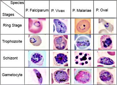
Different Stages of Malarial Parasites Viewed Under Digital Microscope:
How to consistently detect & diagnose cases of malaria both in high & low endemicity regions?
One of the major issues in enabling quick detection of malaria is the lack of technicians in laboratories for microscopic screening of peripheral blood smears. As per government database, there was a shortfall of 12511 lab technicians in PHCs and CHCs across India in 2017, majority in states with high malaria burden such as Orissa, Karnataka, Rajasthan, etc. As a result, blood samples are sent to district hospitals or referral centres for observation, leading to delays of upto 3-7 days in obtaining the result (particularly in remote areas like Gadchiroli district in Maharashtra). This period is very crucial for positive cases leading to fatalities if malaria treatment is not administered within 24 hours. The Strategic Action Plan for Malaria by NVBDCP states that at least 80% of those suffering from malaria should get accurate, affordable, and proper diagnosis within 24 hours of reporting to the health system, which entails that microscopic analysis of blood samples under Digital Microscope or Laboratory Microscope be facilitated at PHC level.
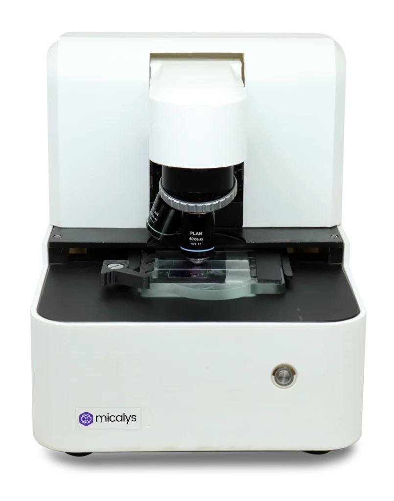
CILIKA Digital Microscope is the ultimate solution for detecting malaria parasite in red blood cells (RBC)!

Malaria parasites are identified by examining a drop of patient’s blood under CILIKA Digital Microscope spread out as ‘blood smear’ on microscope slide. CILIKA microscope has multiple variants like:
CILIKA Portable, world’s first smartphone integrated Digital Microscope with TrueView Technology, is the perfect choice for work-from-home and extensive travelling. Its unmatched high-quality imaging, battery backup and lightweight body with a travel bag makes it the undisputable choice for field visits, medical camps and seminars. Cilika Portable’s 5200 mAhLithium-ion batteries provide 15-hour battery backup, allowing you to travel and work with limited or no power supply. Weighing only 3 kgs, Cilika Portable Digital Microscope fits easily inside a 30 cm X 20cmX 18 cm bag for hassle-free travel, and it comes with a smart business sling bag.
CILIKA Benchtop Series is master blend of traditional and Digital Microscope with features like 4X, 10X, 100X (oil immersion) infinity-corrected achromat lenses, darkfield and phase contrast microscopy, used in diagnostic labs and hospitals for testing samples like blood, semen, tissues, microbes, parasites, smears.
Cilika Transform Series is versatile binocular microscope upgraded to digital benchtop system instantly, apt for students requiring binocular microscopes for classes and examinations.
CILIKA’s TF-100 Digital Trinocular Microscope with universal mobile viewing attachment has hi-tech features like Viewing Head, Eyepiece, Nosepiece, Four High Contrast Infinity Corrected Plan Achromat Objective Lenses with Data Management, Image Capturing, Video Recording and other specifications.
Cilika’s Role in Telepathology for Malaria & Other Diseases – Cilika Digital Microscopes are enabled with best digital setup for telepathology, allowing professionals to live stream whole slides in real-time for second opinions and diagnosis of malaria parasites and other vector-borne diseases and others with pathologists and doctors-capturing, storing in cloud, sharing high-resolution images, videos. Sync CILIKA with Google Drive via CILIKA’s one-touch cloud storage safety, accessing all data across all devices. Click, store, share using WhatsApp, telegram, Gmail, Google Drive, etc. Stream live demonstration on multiple devices simultaneously with CILIKA’s disturbance free multiple device connectivity feature.
How Cilika can be used for malaria diagnosis in rural & urban areas?
Using Cilika Digital Microscope, all PHCs and malaria clinics will be capable of providing correct screening of a blood smear for malaria within 12 hours, regardless of the presence of a trained technician. If technician is absent, telepathology will enable remote diagnosis by technician or pathologist located in another centre/city/hospital. Cross verification of malaria microscopy of positive samples under Cilika Digital Microscope will be performed on regular basis via digital images of smear capture and uploaded by technicians at PHCs/malaria clinics.Purchase CILIKA at the earliest!