

What is Automated Microscopes and its Potential in Decentralizing Pathology?
Conventional microscopes have helped us come a long way. They’ve enabled many pathological studies and supported worthwhile research and studies.
Of course, we acknowledge the contributions of traditional microscopes. But amid the digital age that we live in, we cannot discount new-age needs. The world now demands advanced, intelligent imaging systems that enable quicker, more accurate analyses.
It is here that automated microscopes step in!
Automated microscopes are an evolved form of vintage microscopes. They automate various otherwise arduous, time-consuming, and taxing tasks like observing slides, counting cells and interpreting results. They seem the right fit within the modern context and, thus can be considered a breakthrough innovation.
So, let’s look at some crucial aspects of automated microscopy, like its role in modern labs, how it works and what are its various benefits.
Automated Microscopes in Modern Labs
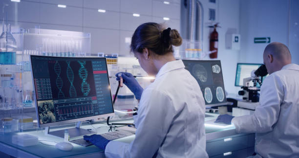
Automated microscopes play a massive role in modern laboratories. Conventional microscopes, governed by out-and-out manual operations, incur a lot of time and effort in adjusting slides, focusing and examining them. Not to mention the factor of human error and subjectivity!
Automating microscopy involves capturing images and automating the overall microscopy workflow. Thus, the pathologist or researcher intervenes in the process only when the system produces the images or churns out results.
It is frequently used in applications that involve repeated observations across an extended timeframe. From live cell imaging to high throughput analysis, automated microscopy enables pathologists and researchers to explore more.
In addition, automating microscopy can help its users scan the entire slide without intervening in the process manually. Furthermore, integrating technologies like AI can allow AI-based intelligent and accurate sample analysis.
How Does Automated Microscopes Work?
Microscopy can be automated in various ways. But the most effective form of automation comes via focus and stage control. The use of focus motors on focus transmission gears can allow image acquisition software to autofocus.
In addition, automation enables the user to select wavelengths, thus enabling users to conduct various experiments, even when the team isn’t physically present in the lab. They can use various elements like acousto-optic tunable filters (AOTFs), beam-splitting units, filters, and monochromators to select wavelengths.
Automated microscopes can also operate in various other ways. Users can automate illumination sources, shutters, image acquisition, and environmental control to increase productivity and enhance microscopy applications.
The most common form of microscopy automation is the whole slide scanner.
Benefits of Automated Microscopes
Microscopy has evolved significantly with time. Digital microscopes and automated microscopes are the latest forms that have revolutionized microscopy while offering several benefits. Convenience is one of the most vital benefits that automated microscopes offer. But primarily, it is the enhanced functional value that they offer that matters more than everything else. Here’s more to it.
- Data Acquisition
Acquiring data in real time is a challenge with manual microscopes. It is because pathologists either would require help or wait until the slide examination is over to acquire the required data. But automated or digital microscopes allow them to fetch data in real-time and in a format that enables quicker interpretation and decision-making.
- Real-Time Analysis
Automated microscopes enable AI-based analysis. It helps reduce manual analytical errors and fosters more accurate and data-driven evaluation. While AI based software cannot replace the expertise of a skilled pathologist, they can go a long way in assisting them, and reducing the time taken for diagnosis by highlighting potential areas of significance or performing repetitive tasks such as cell counting. The combination of technology and human expertise can minimize the error rate significantly.
- Instant Archival
With digital or automated microscopes at the user’s disposal, they can instantly archive data stored virtually on the cloud. Users can access documents without scanning through physical records, thus saving a lot of time and effort. While expediting decision-making, cloud-based quicker and simpler document access helps keep records, observations, analysis, etc., secure.
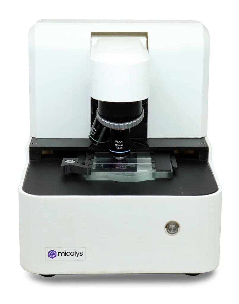
MedPrime Ventures Into Microscopy Automation
While MedPrime with its range of Cilika digital microscopes including the portable, benchtop and upgradeable binocular variants have created an impact in the growth of digital microscopy, automation of microscopy has always been its end goal. The development of its latest innovation MICALYS, has been going on behind the scenes for a few years now. The launch of this exciting new product with the potential to revolutionize the pathology and diagnostics segment is coming soon, and everyone is invited to take a sneek-peek. Stay tuned to our social medial pages (Facebook) for more news on MICALYS!