

Digital Microscope for Education: Better Than Old Microscopes?
In medical training, the tools we use to teach the next generation of physicians are as critical as the curriculum itself. For decades, the traditional optical microscope has been the cornerstone of histology and pathology labs. However, as technology advances, medical institutions are shifting toward more sophisticated alternatives. A digital microscope for education is redefining how students learn, collaborate, and master the intricate details of biological specimens. But is this modern tool truly superior to the classic instruments we have relied on for generations?
The Role of Microscopy in Medical Education
Microscopy is the gateway to understanding cellular biology, pathology, and diagnostic medicine. For an MBBS student, the ability to identify tissue structures, recognize cellular abnormalities, and correlate findings with clinical presentations is paramount. Traditionally, medical colleges have invested heavily in rows of monocular or binocular optical microscopes. While these devices provide the foundational skill of manual focusing and specimen navigation, the pedagogical approach in a modern microscope for medical college needs to be more dynamic, collaborative, and evidence-based.
The primary goal of medical education is to ensure that every student achieves diagnostic accuracy and retains complex visual information. As we embrace digital transformation, the digital microscope for education is proving to be a vital asset in meeting these learning outcomes.
Challenges of Teaching with Conventional Microscopes
Despite their historical importance, conventional optical microscopes present significant hurdles in a modern classroom setting. Because only one student can view a specimen at a time through the eyepiece, instructors spend an inordinate amount of time moving from student to student to verify if they are looking at the correct field of view.
Furthermore, these older systems do not support modern collaborative learning. If a rare pathology slide is being examined, the instructor cannot easily show it to the entire class simultaneously. This often leads to student frustration and inconsistent learning experiences. When searching for the best digital microscope for medical students, educators often highlight these exact pain points—the lack of documentation, the inability to archive findings, and the physical strain associated with long hours at the eyepiece.
What Makes a Digital Microscope Better for Education?
The transition to a digital microscope for education offers a paradigm shift in how histology and pathology are taught. By bridging the gap between physical specimen observation and digital data processing, these systems enhance every aspect of the learning journey.
Live Screen Projection for Group Learning
One of the most transformative features of a digital system is the ability to project live images onto large screens or smartboards. This turns a microscope into a collaborative learning tool. The instructor can guide a lecture, pointing out specific cellular structures while the entire room follows along in real-time. This ensures that every student, regardless of their position in the lab, has the same high-quality visual reference.
Record & Replay for Asynchronous Study
In a high-pressure environment like an MBBS program, the ability to review material at one’s own pace is invaluable. A digital microscope for MBBS students allows for high-resolution image and video capture. Students can record the slide as they navigate it, creating a digital library of specimens to revisit during exam preparation. This feature transforms passive observation into an active, long-term learning resource.
Remote Demonstrations for Online & Hybrid Classes
Modern medical education is no longer confined to the physical laboratory. The rise of hybrid learning models requires tools that function just as effectively remotely as they do in person. A digital microscope for education can be integrated with video conferencing platforms, allowing professors to stream microscopic views directly to students’ devices at home. This continuity is essential for modern curriculum delivery.
Consistent View for Every Student
The best digital microscope for medical students ensures uniformity. In a room of 50 students, each person sees the exact same image with the same lighting and focus. This eliminates the variability of manual focus quality and ensures that every learner is observing the intended pathology without needing to repeatedly ask the instructor for verification.
Digital Microscope vs Old Microscope: Side-by-Side Comparison
When debating digital vs optical microscope education, it helps to look at the tangible differences.
| Feature | Conventional Optical Microscope | Digital Microscope |
| Viewing | Monocular/Binocular (One person) | Live Screen/Display (Group/Class) |
| Documentation | None (Mental/Manual Notes) | Digital Files |
| Collaboration | Low | High (Sharing/Streaming) |
| Physical Strain | High (Eye/Neck fatigue) | Low (Ergonomic viewing) |
| Integration | Standalone | Integrated with E-learning |
The digital vs optical microscope education comparison clearly favors digital systems for their ability to integrate into the modern, data-driven medical curriculum. While the optical microscope remains a classic, the digital version is arguably the best digital microscope for medical students who need to interface with data as much as physical tissue.
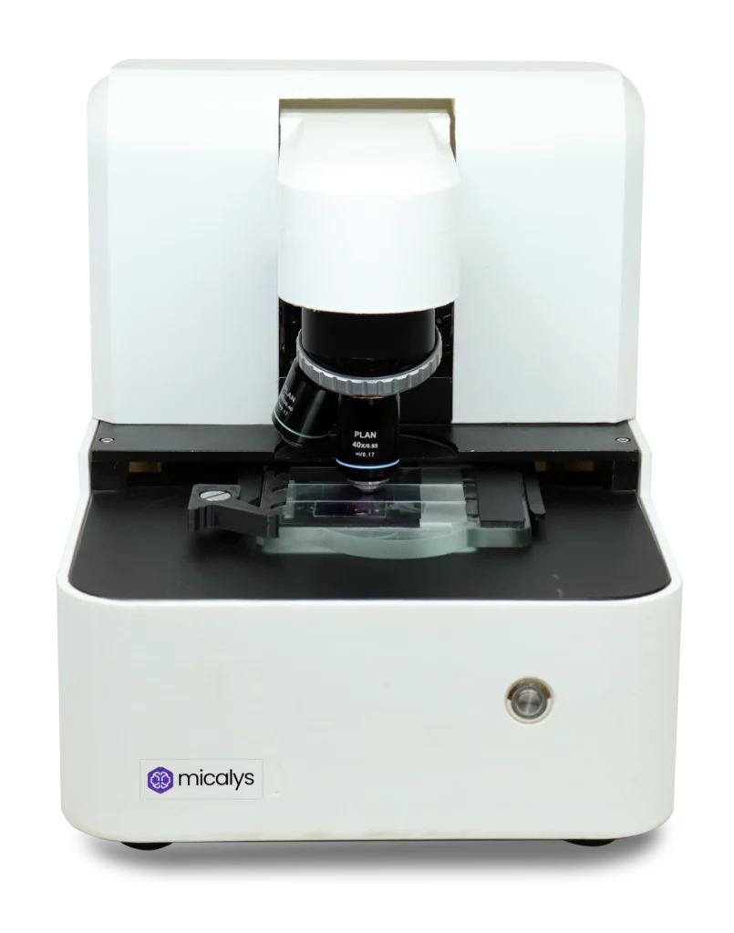
Real Examples: How Indian Medical Colleges Are Using CILIKA
MedPrime Technologies has been at the forefront of this shift. Through our CILIKA range, we have seen various Indian medical colleges adopt digital solutions to modernize their labs. Institutions have integrated CILIKA systems into their pathology department to allow students to capture high-quality digital images of slides, which are then compiled into a digital atlas. This setting leads to a measurable increase in student engagement. By investing in the right microscopes, these institutions are not just buying hardware; they are upgrading their entire diagnostic teaching framework.
Things to Consider Before Buying for Educational Use
Choosing the right digital microscope for MBBS training is a strategic decision. Consider these factors:
- Image Quality & Resolution: Ensure the sensor provides crisp, clear images that accurately represent the tissue stain.
- Ease of Integration: The system should work seamlessly with existing computers, projectors, and learning management systems.
- Durability: A microscope for medical college use must withstand the daily wear and tear of a bustling laboratory environment.
- Software Capability: Does the accompanying software allow for annotation, measurement, and easy file organization?
- Support and Service: Opt for a digital microscope that offers robust local support and training, ensuring that faculty can make the most of the technology.
The debate between the traditional and the modern is common in medicine, but the evidence is clear: the digital microscope for education is not merely a fancy upgrade—it is a functional necessity for the modern medical student. While traditional optical microscopes taught us the basics, the digital era requires tools that facilitate better collaboration, efficient documentation, and hybrid teaching.
By integrating the best digital microscope for medical students—such as those offered by MedPrime Technologies—into the classroom, medical colleges are better preparing their students for a digital-first medical future. If you are ready to modernize your histology or pathology lab, the time to transition to a digital microscope for MBBS students is now. Embrace the change, enhance your teaching, and elevate the learning experience for every student in your care.