


Whole Slide Scanner
Micalys is an automated digital microscope and whole slide scanner, that delivers advanced imaging and telepathology solutions for hospitals, laboratories, and educational institutions.
Whole slide imaging at 100x objective, Z-stacking capabilities, and enhanced telepathology with Quick Share, Secure IP Sharing, and Live View with Robotic Remote Control
Trusted by experts, Micalys transforms whole slide imaging into an effortless, high-precision experience
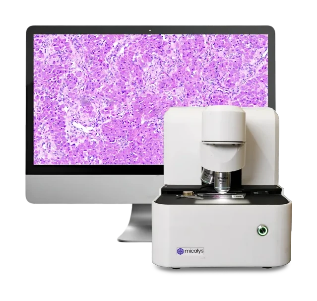
Get to Know Micalys
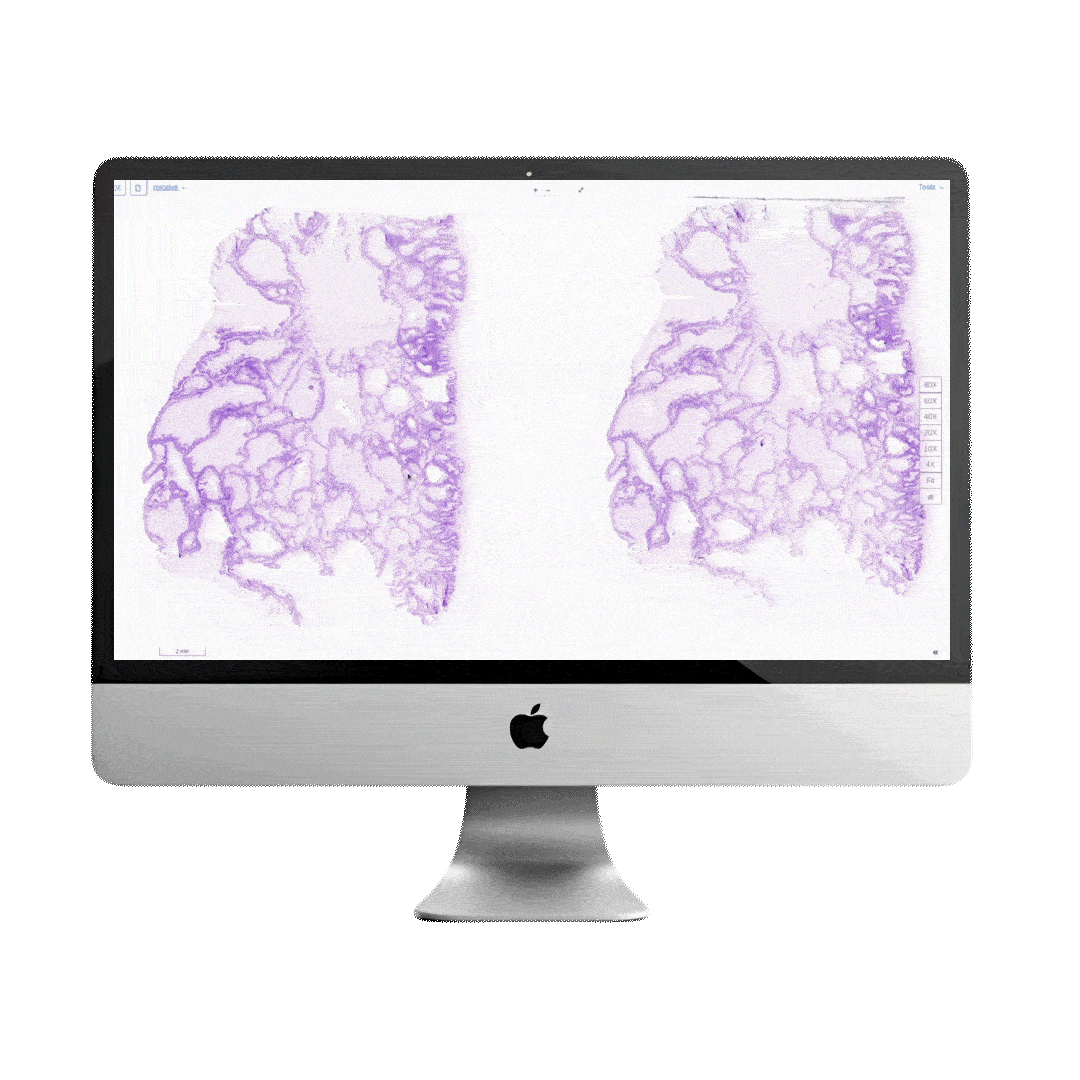
WSI at 100x objective
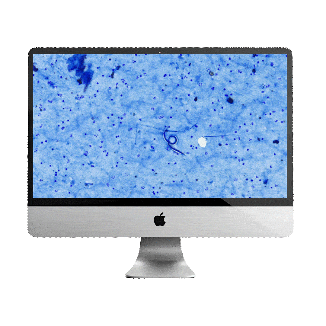
Beyond standard 20x and 40x magnification, this scanner features true 100x oil immersion magnification, providing unparalleled clarity and detail.
Perfect for studying microbiology samples, it allows you to observe every structure closely and effectively.
High resolution Imaging
High-resolution imaging with fast scanning time enables rapid, precise diagnoses in pathology.
This technology minimizes misdiagnoses, reduces the need for repeated tests, and supports high throughput for quicker reporting.
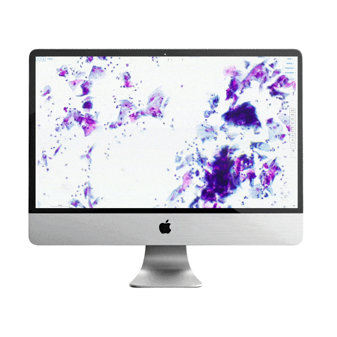
11 layers of z stacking
No need to worry if the slides are uneven, poor quality, or thick. With 11 layers of Z-stacking, the scanner can capture multiple focal planes for clear, detailed imaging.
This feature ensures you gain insights from every layer, leaving no room for information loss. It’s especially useful for cytology samples.
Boost to telepathology Second opinions made easy
Quick Share: Generates a link for easy sharing, perfect when you need a second opinion from a colleague or want to quickly share a case for review—no complications, just fast and simple
Secured IP Sharing: Allows you to securely and easily share findings with your team. Ideal for teams that prefer online diagnosis, ensuring safe and seamless collaboration.
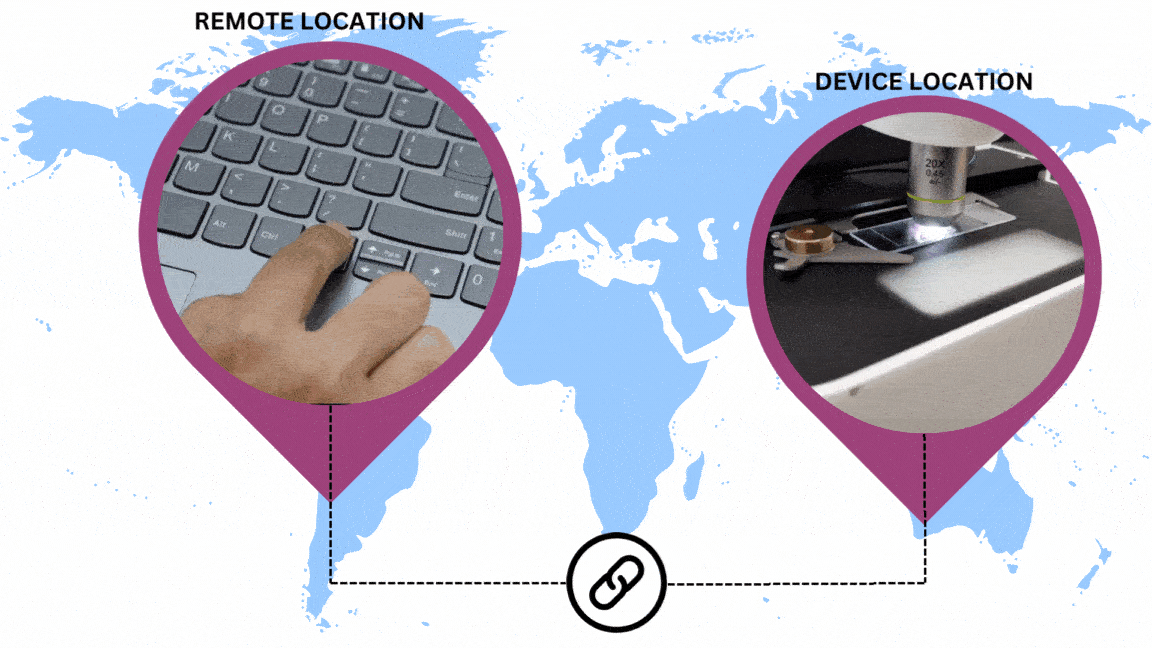
Live view with robotic remote control
More than just a live view, Micalys lets you remotely manage the microscope’s stage, and focus, and provides magnification with an astonishing latency of just 250 milliseconds.
This allows you to work anytime, anywhere, without the need to physically carry the microscope or be present in your lab.
Manual Microscopy
Use the same microscope as a regular digital microscope with the joystick
Helpful when WSI can’t be performed on non-fixed samples like urine, sperm, or stool, or when the sample is damaged
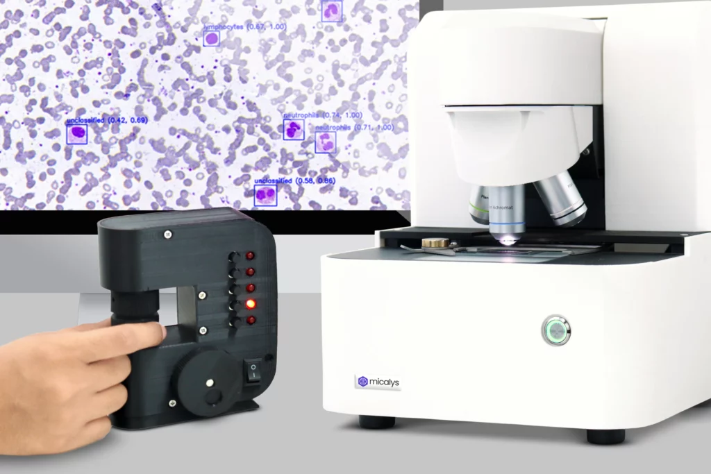
AI Assistance
AI-assisted diagnosis for PBS, Malaria, and Filaria provides advanced support in detecting and analyzing samples
The integration of AI ensures future-ready capabilities, keeping diagnostic processes aligned with the latest advancements.

Why MICALYS - A complete package

Data storage and management
Micalys has its data management software and does not require any additional software 1TB local storage, expandable up to 4TB, along with a network-attached storage (NAS) system for on-premises backup, providing the desired storage space

User-friendly interface
The clean interface and minimalist design make it easy for both doctors and lab technicians to adopt, ensuring a smooth and efficient experience

Compact and Robust design
The robust design prevents device malfunctions and calibration issues, ensuring accurate results and minimizing downtime for consistent performance
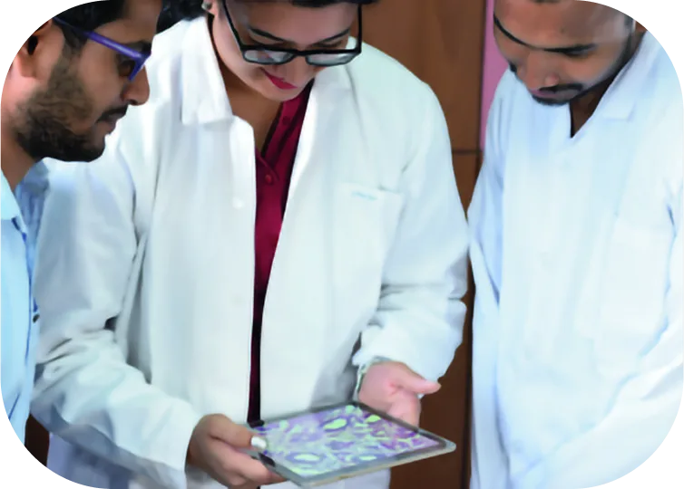
Testimonials
Product Highlight


Ready to experience the future of microscopy?
Book a personalized demo; either in person with your sample or online in just 2 minutes
Schedule Demo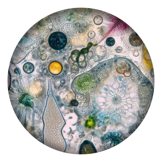
FAQ
What are the sample types that can be observed under Micalys?
Using micalys, one can perform whole slide imaging on,
-
- Histopathology
- IHC
- Cytology
- Blood
What resolution can be achieved with a Micalys scanner?
The resolution depends on the objectives used, ranging from 20x to 100x magnification.
-
- 20X: ~0.50 µm/pixel
- 40X: ~0.25 µm/pixel
- 100X: ~0.11 µm/pixel
What is the difference between Micalys S1, M1, and M3
Micalys S1: It has a single slide capacity and one lens hole
Micalys M1: It has a single slide capacity and four lens holes
Micalys M3: It has triple slide capacity and four lens holes
What is Z-stacking, and how is it helpful?
Z-Stacking is the process of capturing multiple images of a slide at different focal depths and stacking them together to create a composite image
Micalys offers 11 layers of Z stacking
Benefits:
Improves focus across uneven or thick samples
Essential for cytology samples
What software is required to view digital slides?
Micalys offers its web-based solution for viewing and analysis.
Is it possible to share scanned slides with colleagues remotely?
- Yes, Micalys enables you to share digital slides via various methods as per the requirements, enabling remote access and collaborative review.
- Quick share: Share the scanned image of the whole slide in digital format via a quick shareable link
- Secured IP sharing: Create a secured group in the software with relevant team members. Share the scanned image, and everyone can view and share their opinions from their respective locations
- Certainly, you may capture quick screenshots and share as PNGs, or share your screen effortlessly in an online meeting on GMeet, Zoom, or Teams