

How Digital Microscopes Are Transforming Pathology Reporting
Precision and speed are the cornerstones of patient care. For decades, experts have relied on conventional optical tools to interpret tissue samples. However, the paradigm is shifting. Today, the integration of a digital microscope in every pathology lab is fundamentally changing how experts approach reporting. As hospitals and diagnostic centers move toward digitization, the modern digital microscope has become an essential tool for pathology workflows, ensuring that clinical reporting is not only faster but also significantly more accurate than ever before.
The Traditional Pathology Reporting Challenge
Traditional pathology has long relied on physical glass slides viewed through an analog ocular. While this method served the medical community well for over a century, it is fraught with manual inefficiencies that directly impact reporting timelines. Pathologists face physical limitations, such as the need to be physically present at the lab to view samples, which inevitably creates a bottleneck.
Furthermore, manual documentation in reporting—where a pathologist dictates or types findings based on what they see through a traditional microscope – is prone to human error. The lack of standardized digital storage makes retrieving historical slides difficult, and the subjective nature of manual slide examination can lead to inter-observer variability. When every minute matters for a patient awaiting a diagnosis, these traditional workflows often fail to meet the high-throughput demands of modern clinical pathology and reporting requirements.
What Has Changed with Digital Microscopy?
The transition to a high-quality digital microscope has bridged the gap between traditional microscopy and the digital age. By capturing high-resolution images of tissue samples, these systems allow experts to analyze specimens on large, high-definition screens, reducing physical strain and enhancing diagnostic clarity in pathology reporting.
Whole Slide Imaging (WSI)
Whole slide imaging is the backbone of this transformation. Instead of focusing on small fields of view, whole slide imaging allows the entire specimen to be digitized into a single, navigable file. This technology enables pathologists to zoom in and out with ease, mimicking the experience of a traditional digital microscope but with the added benefit of digital annotation and measurement tools that refine the pathology reporting process.
AI-Powered Diagnostic Support
The integration of Artificial Intelligence (AI) with a digital microscope has opened new frontiers. AI algorithms can now perform initial screenings, highlighting suspicious regions of interest (ROI) that the human eye might overlook. This speeds up pathology review, allowing the pathologist to focus their expertise on high-stakes analysis, ultimately improving the quality of the final pathology reporting.
Cloud-Based Sharing & Secure IP Access
Modern pathology requires seamless collaboration. Telepathology solutions leverage the cloud to allow instant sharing of high-resolution slide data captured by a digital microscope. Whether for a second opinion or a multidisciplinary team meeting, a digital microscope connected to a secure network ensures that expert input is just a click away, making it a critical component of digital pathology India’s rapidly modernizing infrastructure for clinical reporting.
How Digital Microscopes Speed Up Report Turnaround
Efficiency in reporting is tied directly to the workflow speed provided by a digital microscope. By eliminating the need to physically handle and transport glass slides, labs can process samples faster. Digital workflows allow for seamless integration with Laboratory Information Systems (LIS). When a pathologist uses a digital microscope to finalize findings, the data can be automatically pushed into the reporting module, reducing administrative burdens and minimizing the time from sample reception to final pathology reporting.
Remote Pathology: Diagnosing Across Distance
Geographical barriers have historically limited access to specialized diagnostic expertise. Telepathology solutions have demolished these barriers. Today, a digital microscope in a small, rural clinic can send high-fidelity images to a super-specialist in a metropolitan hospital. This accessibility is a game-changer for digital pathology India, ensuring that high-quality pathology reporting is available regardless of location, supported by the precision of a modern digital microscope.
Real-World Impact: Labs Using Digital Microscopes
Diagnostic centers that have adopted a digital microscope approach have reported a marked improvement in their daily operations. By shifting to a digital-first pathology workflow, these labs have seen a significant decrease in turnaround time. The ability to digitally archive cases also simplifies audit processes and teaching requirements, demonstrating that the best microscope for pathology reports is one that is fully integrated into a digital ecosystem to streamline reporting.
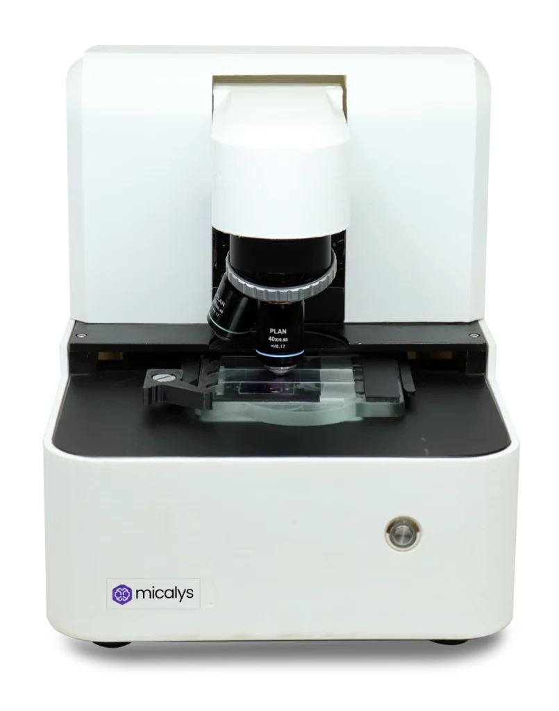
MICALYS & CILIKA: Medprime’s Role in This Transformation
Medprime Technologies is at the forefront of this revolution. With our flagship products like CILIKA and MICALYS, we are providing the best microscope for pathology reports that balances affordability with high-end performance. Our solutions are designed to address the specific needs of digital pathology in India, offering robust whole slide imaging capabilities and seamless connectivity. By combining hardware excellence with user-centric software, Medprime ensures that every digital microscope we deploy helps pathologists create a more precise and efficient pathology reporting experience.
What to Expect Next in Digital Pathology
We expect to see more compact digital microscope systems that can process complex slides in real-time. As telepathology solutions become more sophisticated, the speed and security of reporting will continue to improve, making advanced diagnostic tools accessible even in the most remote corners of the globe through a digital microscope.
The shift toward the digital microscope is not merely a technological upgrade; it is a fundamental improvement in patient care. By enhancing the efficiency of pathology and standardizing the quality of reporting, these tools are empowering doctors to make faster, more accurate decisions. For those seeking the best microscope for pathology reports, Medprime Technologies remains committed to driving innovation in the sector. As we continue to embrace digital transformation, the promise of a more connected, efficient, and precise world of pathology reporting becomes an increasingly tangible reality.